|
Medical Imaging Interaction Toolkit
2016.11.0
Medical Imaging Interaction Toolkit
|
|
Medical Imaging Interaction Toolkit
2016.11.0
Medical Imaging Interaction Toolkit
|
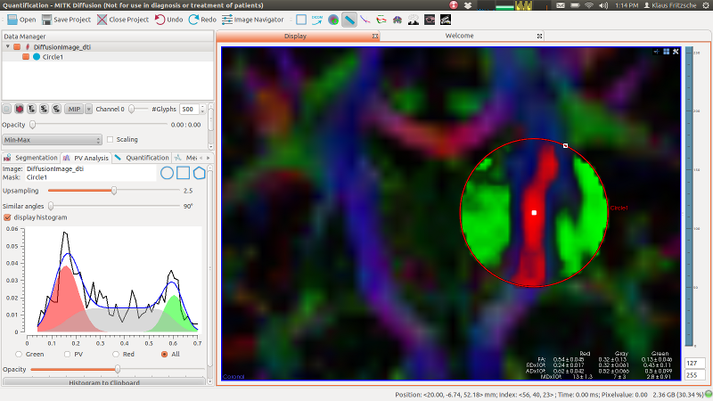
The "Partial Volume Analysis" view can be found in the "Quantification" perspective. It allows for robust quantification of diffusion or other scalar measures in the presents of two classes (e.g. fiber vs. non-fiber) and partial volume between them. The algorithm estimates a probabilistic segmentation of the three classes and returns a weighted average of the measure of interest within the each class.

All measures are automatically written to the clipboard once the estimation is updated. For scalar images, the mean and the variance of the gaussian selected in the radio-box above is copied out. If 'All' is selected, then all means and variances are carried out as a tab-separated list. For tensor images, the values for all scalar measures (FA, MD, RD and AD) are carried out to the clipboard.
The histogram export is provided by the button underneath the histogram. The values can be pasted to excel or any text editor.
Are not recommended for use yet.
Diffusion tensor imaging in primary brain tumors: reproducible quantitative analysis of corpus callosum infiltration and contralateral involvement using a probabilistic mixture model. Stieltjes B, Schlüter M, Didinger B, Weber MA, Hahn HK, Parzer P, Rexilius J, Konrad-Verse O, Peitgen HO, Essig M. Neuroimage. 2006 Jun;31(2):531-42. Epub 2006 Feb 14. PMID: 16478665